A modern approach to cataract surgery
Cataract is the opacification of the crystalline lens and one of the leading causes of reversible vision loss worldwide. The surgical indication is clinical and considers the functional impact reported by the patient, not merely the examination findings.

Clinical explanation
Preoperative evaluation includes refraction, biomicroscopy, tonometry, retinal mapping and optical biometry for intraocular lens calculation. Corneal topography assists in the analysis of astigmatism.
Phacoemulsification is the reference technique: through small incisions, the opacified lens is fragmented and aspirated, followed by implantation of an intraocular lens within the capsular bag.
Lens selection takes into account the patient's lifestyle, corneal astigmatism, and the condition of the retina and optic nerve. Monofocal, toric and extended-depth-of-focus lenses have distinct indications, discussed on a case-by-case basis.
Clinical timeline
Stages of care
- 01
Consultation and indication
Assessment of the functional impact of the opacity and a complete ophthalmologic examination.
- 02
Preoperative testing
Optical biometry, corneal topography and macular OCT when associated retinal disease is suspected.
- 03
Intraocular lens selection
Discussion of the available options, with their advantages and limitations, aligned with the patient's expectations.
- 04
Surgery and follow-up
An outpatient procedure under topical anesthesia, with scheduled re-evaluations and a new refraction after stabilization.
Diagnostic imaging
Imaging documentation
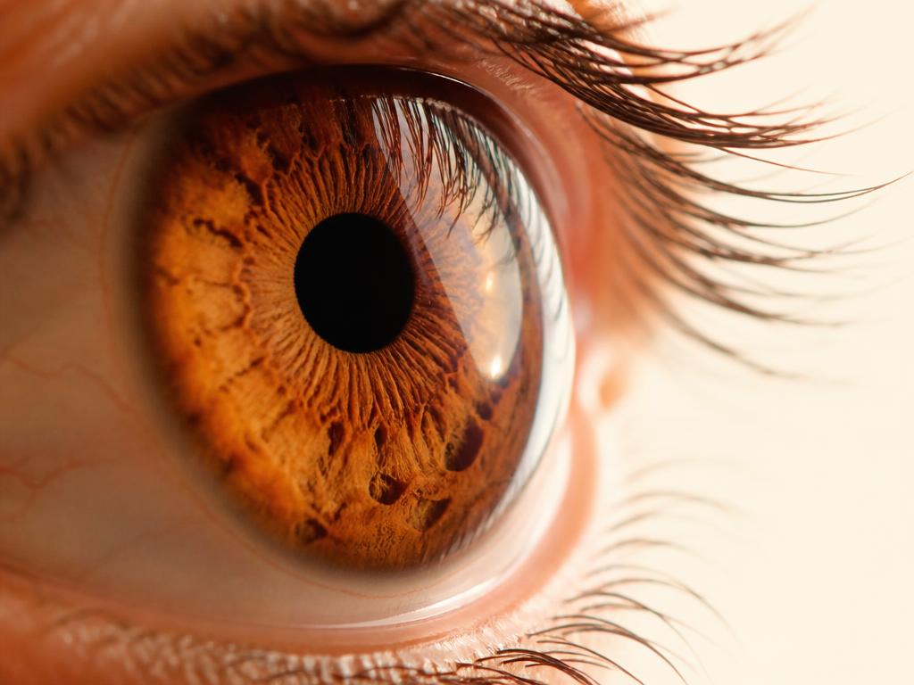
Corneal imaging Assessment of the anterior segment by topography, tomography and specular microscopy, as clinically indicated.
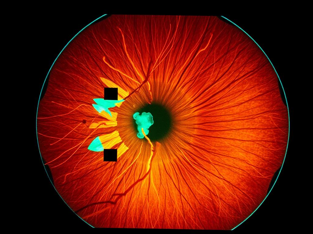
OCT Optical coherence tomography: a high-resolution cross-sectional image of the retinal layers, essential for macular assessment and quantitative follow-up.
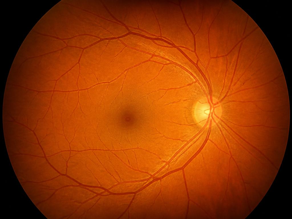
Fundus photography Color documentation of the ocular fundus, used to record the state of the retina and compare its evolution throughout follow-up.
Discussion
Refractive predictability depends on the quality of the biometry and the calculation formula used; small residual variations are expected and described in the literature.
Posterior capsule opacification is a common late event that is treatable in the office with YAG laser capsulotomy.
Expected outcome
Removal of the opacity and visual rehabilitation consistent with the health of each patient's retina and optic nerve are expected. Associated ocular conditions may limit the visual gain achieved.
Related cases
- Retina
Surgical treatment of retinal detachment
An educational overview of the urgent evaluation, the available surgical techniques and postoperative follow-up.
Read the case - Refractive Surgery

Personalized correction for myopia and astigmatism
Eligibility criteria, corneal testing and the different strategies for correcting refractive error.
Read the case - Cornea
Evaluation and management of corneal disease
Keratoconus, dystrophies and corneal opacities: imaging diagnosis and the therapeutic options described in the literature.
Read the case