Surgical treatment of retinal detachment
Retinal detachment is an urgent condition in which the neurosensory retina separates from the retinal pigment epithelium. The interval between diagnosis and treatment is one of the most extensively studied factors in the literature regarding functional prognosis.
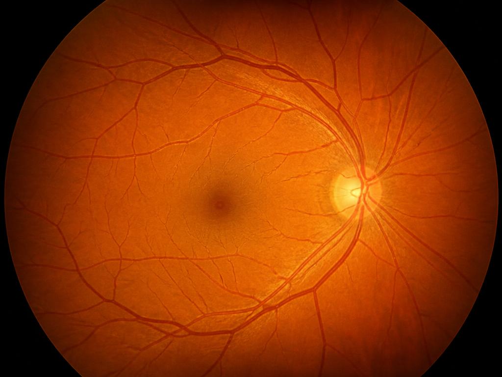
Clinical explanation
The most frequent presentation is the rhegmatogenous form, associated with a retinal tear that allows fluid to pass into the subretinal space. Symptoms such as a sudden increase in floaters, flashes of light and loss of peripheral visual field warrant immediate evaluation.
Dilated fundus examination, complemented by photographic documentation and, when the media do not allow adequate visualization, ocular ultrasonography, defines the extent of the detachment and macular involvement.
The surgical approaches described in the literature include pneumatic retinopexy, scleral buckling and pars plana vitrectomy, used alone or in combination. The choice takes into account the type and location of the tears, the status of the lens and the presence of proliferative vitreoretinopathy.
Clinical timeline
Stages of care
- 01
Urgent evaluation
Directed history-taking, visual acuity measurement, biomicroscopy and dilated retinal mapping.
- 02
Imaging documentation
Fundus photography and, when indicated, ocular ultrasonography and macular OCT to define macular involvement.
- 03
Surgical planning
Discussion of the indicated technique, alternatives and risks, with documentation of informed consent.
- 04
Postoperative follow-up
Serial re-evaluations with fundus examination and OCT, monitoring retinal reattachment and functional recovery.
Diagnostic imaging
Imaging documentation
Fundus photography Color documentation of the ocular fundus, used to record the state of the retina and compare its evolution throughout follow-up.
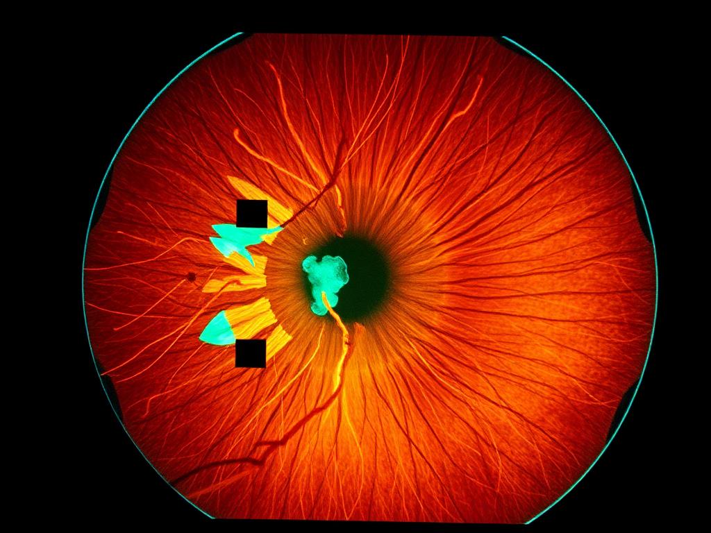
OCT Optical coherence tomography: a high-resolution cross-sectional image of the retinal layers, essential for macular assessment and quantitative follow-up.

Angiography Study of the retinal circulation, indicated when it is necessary to map areas of ischemia or vascular leakage.
Discussion
The vitreoretinal literature describes variable primary reattachment rates depending on technique and case complexity, with the possibility of reoperation in a proportion of patients.
Macular involvement prior to surgery is the principal determinant of visual recovery described in the studies, which underscores the value of early diagnosis.
Expected outcome
The therapeutic goal is anatomical reattachment of the retina and preservation of the visual function possible for each case. Results are individual and depend on the duration of the detachment, its extent, and macular involvement.
Related cases
- Cataract

A modern approach to cataract surgery
How biometry, intraocular lens calculation and phacoemulsification structure contemporary surgical planning.
Read the case - Refractive Surgery

Personalized correction for myopia and astigmatism
Eligibility criteria, corneal testing and the different strategies for correcting refractive error.
Read the case - Cornea

Evaluation and management of corneal disease
Keratoconus, dystrophies and corneal opacities: imaging diagnosis and the therapeutic options described in the literature.
Read the case