Evaluation and management of corneal disease
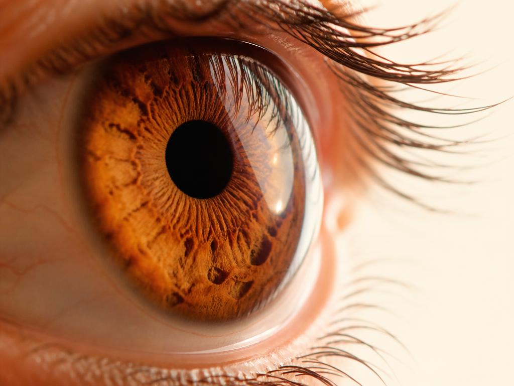
The cornea accounts for most of the eye's refractive power. Changes in its transparency or curvature directly affect visual quality, even with an intact retina.
Clinical explanation
Keratoconus is a progressive ectasia characterized by thinning and irregular steepening. Early diagnosis relies on corneal tomography and serial follow-up.
Corneal collagen crosslinking is the procedure described for stabilizing documented progression, without a primarily refractive purpose.
In advanced opacities, lamellar or penetrating transplantation techniques are considered according to the affected layer, preserving healthy tissue whenever possible.
Clinical timeline
Stages of care
- 01
Surface examination
Biomicroscopy, tear film assessment and photographic documentation of the anterior segment.
- 02
Corneal imaging
Topography, elevation-based tomography, pachymetry and specular microscopy as suspected.
- 03
Treatment definition
Clinical monitoring, stabilization or surgical indication, according to documented progression.
- 04
Visual rehabilitation
Optical fitting and long-term follow-up of corneal transparency and regularity.
Diagnostic imaging
Imaging documentation
Corneal imaging Assessment of the anterior segment by topography, tomography and specular microscopy, as clinically indicated.
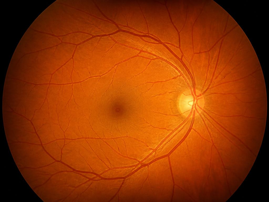
Fundus photography Color documentation of the ocular fundus, used to record the state of the retina and compare its evolution throughout follow-up.
Discussion
Comparison of serial examinations is the accepted method for characterizing progression, avoiding interventions based on a single measurement.
In transplants, prolonged monitoring of the endothelium and the immune response is part of standard care.
Expected outcome
The aim is to preserve corneal transparency and regularity and, when indicated, to stabilize progression. Visual recovery depends on the affected layer and the stage at the time of diagnosis.
Related cases
- Retina
Surgical treatment of retinal detachment
An educational overview of the urgent evaluation, the available surgical techniques and postoperative follow-up.
Read the case - Cataract

A modern approach to cataract surgery
How biometry, intraocular lens calculation and phacoemulsification structure contemporary surgical planning.
Read the case - Refractive Surgery

Personalized correction for myopia and astigmatism
Eligibility criteria, corneal testing and the different strategies for correcting refractive error.
Read the case