Personalized correction for myopia and astigmatism
Refractive surgery encompasses procedures intended to reduce dependence on glasses and contact lenses. Eligibility is determined by objective safety criteria, and not every patient is a candidate.

Clinical explanation
Evaluation includes static and dynamic refraction, corneal topography and tomography, pachymetry, tear film analysis and retinal mapping.
Documented refractive stability and the absence of signs of corneal ectasia are prerequisites discussed in the literature for indicating corneal tissue ablation.
When corneal parameters are unfavorable, alternatives such as phakic intraocular lenses may be considered, always with explicit discussion of the risks and limitations involved.
Clinical timeline
Stages of care
- 01
Refractive screening
Confirmation of refractive stability and review of family history of keratoconus and ocular surface disease.
- 02
Corneal study
Topography, tomography and pachymetry to analyze curvature, thickness and regularity.
- 03
Shared decision-making
Presentation of the techniques indicated for the profile analyzed, with realistic expectations and risks described.
- 04
Follow-up
Scheduled re-evaluations with refraction and ocular surface examination during the first year.
Diagnostic imaging
Imaging documentation
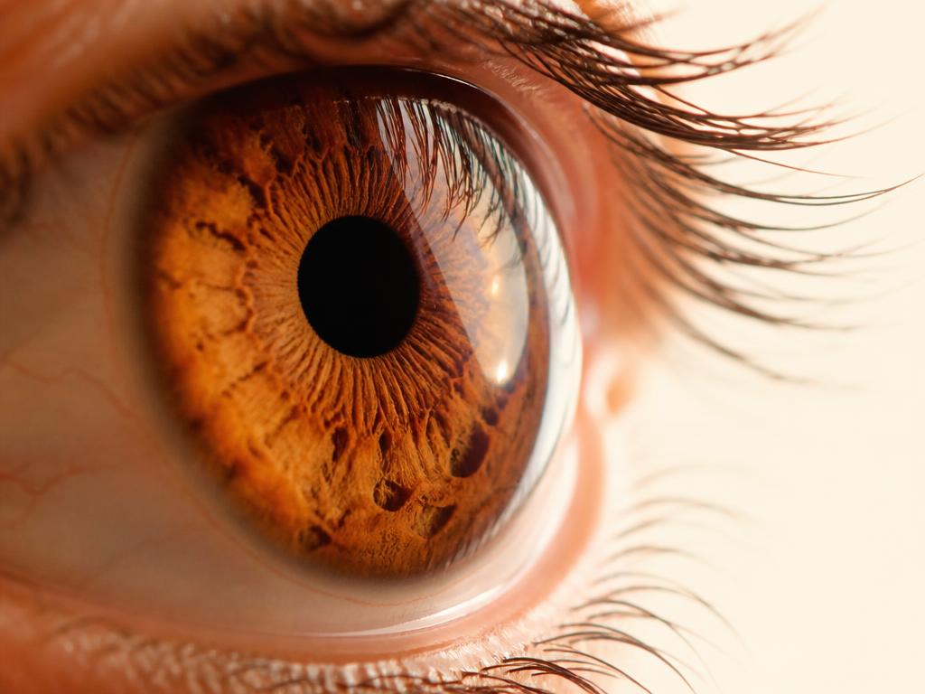
Corneal imaging Assessment of the anterior segment by topography, tomography and specular microscopy, as clinically indicated.
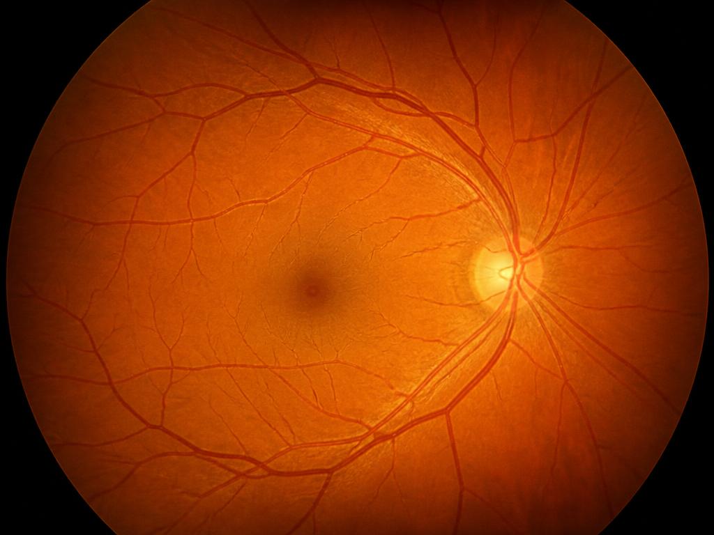
Fundus photography Color documentation of the ocular fundus, used to record the state of the retina and compare its evolution throughout follow-up.
Discussion
Transient symptoms of dry eye and perception of nighttime halos are described in the literature during the early postoperative period and should be disclosed beforehand.
Careful screening for ectasia is the principal safety factor reported in published series.
Expected outcome
The goal is to reduce the refractive error within a range that is safe for each cornea. Partial regressions and occasional need for residual optical correction are recognized possibilities.
Related cases
- Retina
Surgical treatment of retinal detachment
An educational overview of the urgent evaluation, the available surgical techniques and postoperative follow-up.
Read the case - Cataract

A modern approach to cataract surgery
How biometry, intraocular lens calculation and phacoemulsification structure contemporary surgical planning.
Read the case - Cornea
Evaluation and management of corneal disease
Keratoconus, dystrophies and corneal opacities: imaging diagnosis and the therapeutic options described in the literature.
Read the case